OPEN ACCESS: https://rdcu.be/c8xQ5 OR doi.org/10.1007/s10043-023-00792-1
Introducing the SD-OCT: A new tool for visualizing and quantitatively analyzing the dynamics of water content in biological tissue.
- Measure the amount of water localized in biological tissue
- Visualize natural phenomena that have never previously been observed
- Useful for applications in cross-sectional imaging and quantitative analysis of evaporation
The SD-OCT is a powerful new tool that can help scientists and researchers gain a deeper understanding of the role of water in biological activity. By measuring the dynamics of water content in tissue, the SD-OCT can help to identify and track changes in biological processes.
The SD-OCT is also a valuable tool for medical imaging. By visualizing the distribution of water in tissue, the SD-OCT can help doctors to diagnose and treat a variety of conditions, such as dehydration, edema, and cancer. The SD-OCT is a versatile and powerful tool that has the potential to revolutionize the way we understand and treat biological processes.
- The SD-OCT is a non-invasive and painless procedure.
- It can be used to image a wide variety of tissues, including skin, muscle, bone, and brain tissue.
- The SD-OCT is a relatively new technology, but it is quickly becoming an essential tool for scientists and medical professionals.
Imaging and quantitative analysis of water evaporation process using spectral-domain optical coherence tomography under illumination with two near-infrared wavelengths
Optical Review (2023)Cite this article
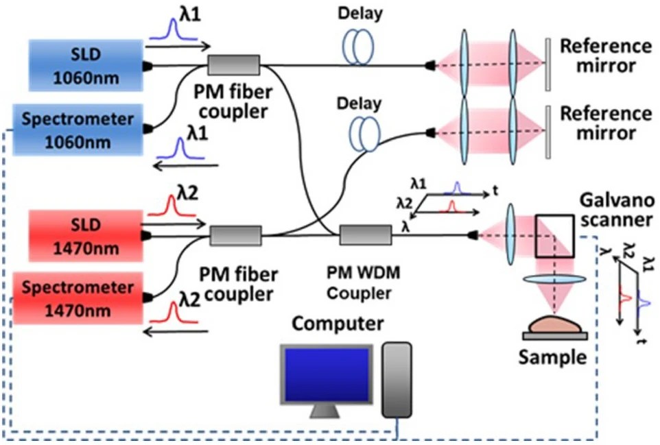
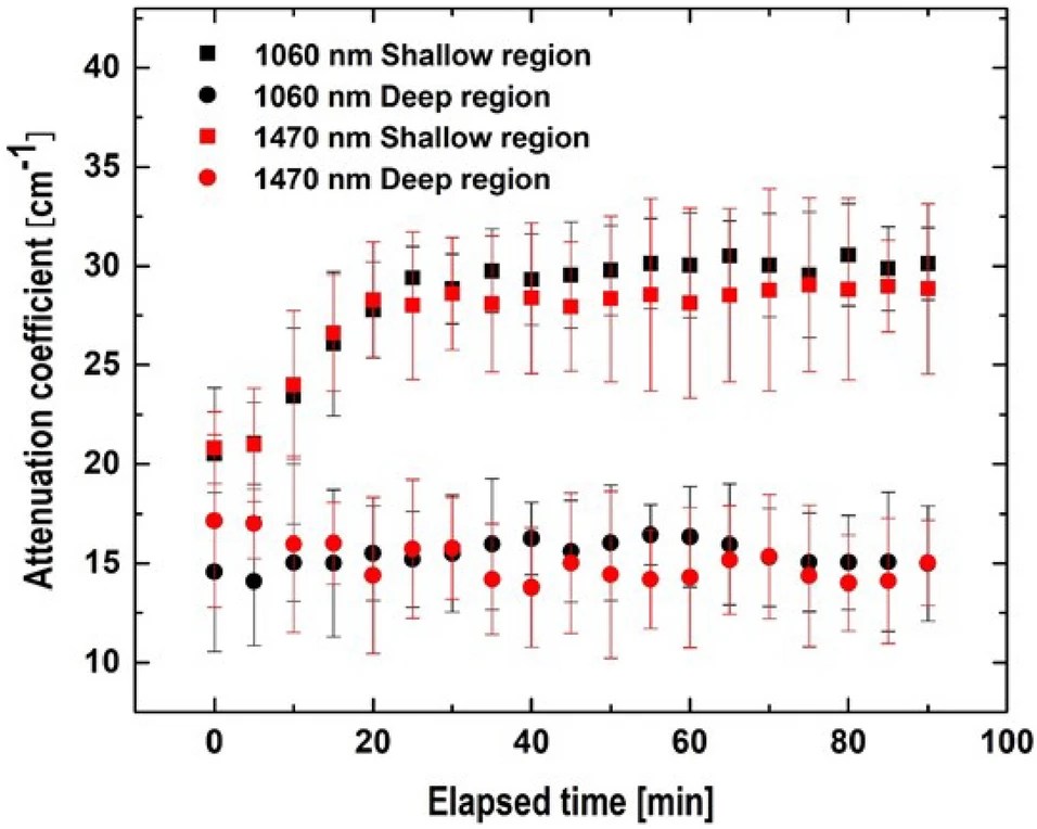
Dynamics of water content in a biological tissue is an important information of a biological activity. However, although the conventional measurement method measures the total amount of water contained in the tissue, there is no established method for quantitatively measuring the amount of water localized in biological tissue. A spectral-domain optical coherence tomography system (SD-OCT) using two near-infrared wavelengths was developed for applications in cross-sectional imaging and quantitative analysis of evaporation at room temperature. The wavelengths of the light sources were selected as 1060 nm, which is hardly absorbed by water, and 1470 nm, which is absorbed by a factor of 220. In this study, we examined the effect of water absorption and scattering on the attenuation coefficient in a dense medium using a two-wavelengths OCT and showed the possibility of measuring the dynamics of water in the evaporation process through light scattering. Experimental results showed that the attenuation coefficient in a dense medium was more affected by scattering than water absorption. They are highly influenced by scattering caused by the temporal variation of the refractive-index matching effect between the hydrogen-bonded cellulose and free water around it in the process of evaporation. It was concluded that the SD-OCT was a quite practical and useful tool to visualize and quantitatively analyze natural phenomena that have never previously been observed.
生体組織中の水分量の動態は、生物活性の重要な情報である。しかし、従来の測定法では組織に含まれる水分の総量を測定するが、生体組織に局在する水分量を定量的に測定する方法は確立されていない。2つの近赤外波長を用いたスペクトル領域光干渉断層計システム (SD‐OCT) を室温での蒸発の断面画像化と定量分析に応用するために開発した。光源の波長は、水にほとんど吸収されない1060 nmと、220倍吸収される1470 nmを選択した。本研究では、2波長OCTを用いて高密度媒質中の減衰係数に対する吸水・散乱の影響を調べ、光散乱による蒸発過程における水の動力学を測定する可能性を示した。実験の結果、高密度媒質中の減衰係数は吸水よりも散乱の影響を強く受けることが分かった。これらは、蒸発の過程で水素結合したセルロースとその周囲の自由水との屈折率整合効果の時間的変動による散乱の影響を強く受ける。SD-OCTは、これまで観測されたことのない自然現象を可視化し、定量的に分析するための非常に実用的で有用なツールであると結論付けられた。
This work was a collaborative study between Iwai Lab and Lenggoro Lab. We are at the same (BASE) building.
Prof. Toshiaki Iwai retired in March 2022. Here is his lab activities’ record.
https://web.tuat.ac.jp/~biome/lab_Iwai.htm